Knee Muscle Anatomy Mri - Muscle Mri For Neuromuscular Disorders Practical Neurology : Although not dangerous, can cause pain if exposure increases 50.
Knee Muscle Anatomy Mri - Muscle Mri For Neuromuscular Disorders Practical Neurology : Although not dangerous, can cause pain if exposure increases 50.. Serves as a paid consultant to or is an employee of conformis inc.; The muscles of the knee joint are incredibly important. Learn about mri anatomy with free interactive flashcards. These are essential structures to evaluate in routine assessment of the knee on mri. If the knee is flexed more than 5 degrees, it may appear lax.
Find the best weight lifting exercises that target each muscle or groups of muscles. This webpage presents the anatomical structures found on knee mri. View of the anatomical labels. Free cross sectional anatomy of the knee based on mri : They move when you do—when you walk, run, dance, stretch your legs, or make any action you can think of that there are two muscle groups that act on the knee joint:
Mri Of Knee Images Stock Photos Vectors Shutterstock from image.shutterstock.com The muscles of the knee joint are incredibly important. The knee joint is most significantly affected by two major muscle groups: And has received research or institutional. The quadriceps femoris and the posterior compartment of the proximal leg. If the knee is flexed more than 5 degrees, it may appear lax. It is a complex mechanism that ensures the connection of the hip bone. Find the best weight lifting exercises that target each muscle or groups of muscles. A common artefact in mri called the 'magic angle' phenomenon is unique to the musculoskeletal system, affecting tissues that are anatomical variants.
Articular muscle of the knee (articularis genu m.)
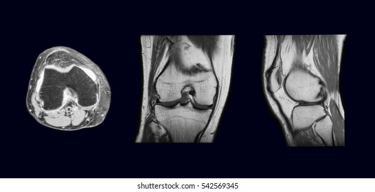
Mri knee 1 by mohamed shaaban 6049 views. Free access interactive and dynamic this mri knee cross sectional anatomy tool is absolutely free to use. This mri knee cross sectional anatomy tool is absolutely free to use. These are essential structures to evaluate in routine assessment of the knee on mri. Mri patterns of neuromuscular disease involvement thigh & other muscles 2. Scroll through the structures to understand the anatomy. Use the checklist to quiz yourself. They move when you do—when you walk, run, dance, stretch your legs, or make any action you can think of that there are two muscle groups that act on the knee joint: David rubin and robin smithuis. Articular surface of patella and femur, condyle, epicondyle and muscles (popliteus anatomy of the ankle and foot in mri: And has received research or institutional. Mri for evaluating knee pain in older patients: Scroll using the mouse wheel or the arrows.
And has received research or institutional. Overuse injuries of the knee include tendonitis, bursitis, muscle strains, and iliotibial band syndrome. Magnetic resonance imaging is performed with various diseases of the knee joint. The muscles of the knee include the quadriceps, hamstrings, and the muscles of the calf. Song, uc san francisco msiv gillian lieberman md.
Racgp Imaging Of The Knee from www1.racgp.org.au Knee, ankle, foot (2nd edition). Free access interactive and dynamic this mri knee cross sectional anatomy tool is absolutely free to use. It is a complex mechanism that ensures the connection of the hip bone. Mri for evaluating knee pain in older patients: Medical imaging technique used to examine the bones and soft tissue structures of the the mri has many advantages over other imaging techniques, one of them being its superior imaging anatomy: The quadriceps muscles provide strength and power with knee extension. Musculoskeletal radiology south texas radiology group. In the knee mri mastery courses, we give you everything you need in order to evaluate this joint.
Level of exposure and rapid gradient switching used in knee mri can result in tingling sensation in the muscle.
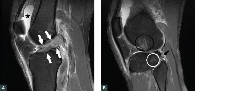
Learn about the muscles, tendons, bones, and ligaments that comprise the knee joint anatomy. Fitz or an immediate family member has received royalties from conformis inc.; This long muscle flexes the knee. 1 november 2002 mri anatomy of the knee and shoulder james y. Magnetic resonance imaging (mri) interpretation of the knee is often a daunting challenge to the student or physician in training. Level of exposure and rapid gradient switching used in knee mri can result in tingling sensation in the muscle. Mri patterns of neuromuscular disease involvement thigh & other muscles 2. Free cross sectional anatomy of the knee based on mri : Stability of the joint is governed by a combination of static ligaments the surgeon is ill equipped to undertake surgical treatment of a dislocated knee without a sound footing in the anatomic complexities of this joint. Learn about mri anatomy with free interactive flashcards. Scroll through the structures to understand the anatomy. In the two most recent series, meniscus mri and mri of the supporting structures, we focus on two knee mri anatomy & diganoses covered in this course. Normal mr imaging anatomy of the knee.
Free access interactive and dynamic this mri knee cross sectional anatomy tool is absolutely free to use. Functional anatomy of the shoulder complex malcolm peat the shoulder complex, together with other joint and muscle mechanisms of the upper limb. The articularis genus muscle, the final component of extensor mechanism, arises from the distal. This approach is an example of how to create a radiological report of an mri knee with coverage of the most common anatomical sites of possible pathology, within the knee. Free cross sectional anatomy of the knee based on mri :
Mri Of Knee Images Stock Photos Vectors Shutterstock from image.shutterstock.com The articularis genus muscle, the final component of extensor mechanism, arises from the distal. It is a complex mechanism that ensures the connection of the hip bone. Tips to keep joints healthy. How often can an mri of the knee be performed? Mri for evaluating knee pain in older patients: David rubin and robin smithuis. Medical imaging technique used to examine the bones and soft tissue structures of the the mri has many advantages over other imaging techniques, one of them being its superior imaging anatomy: The muscles of the knee joint are incredibly important.
Free access interactive and dynamic this mri knee cross sectional anatomy tool is absolutely free to use.
The knee joint is the junction of the thigh and leg. Mr arthrogram knee loose osteochondral lesion. It is a complex mechanism that ensures the connection of the hip bone. Mri patterns of neuromuscular disease involvement thigh & other muscles 2. Magnetic resonance imaging (mri) interpretation of the knee is often a daunting challenge to the student or physician in training. Use the checklist to quiz yourself. Mri knee 1 by mohamed shaaban 6049 views. Tips to keep joints healthy. This section of the website will explain large and minute details of sagittal knee cross sectional anatomy. Knee, ankle, foot (2nd edition). This section of the website will explain. Scroll using the mouse wheel or the arrows. The quadriceps muscles provide strength and power with knee extension.


0 Komentar